On a Blood Smear Slide Prepared Using Wright's Stain
If it is poorly made. Add equal number of drops of buffered water pH 68 on the slide.
Blood Smear Preparation And Staining
Most commercial laboratories use some form of Romanowsky-type stain eg.

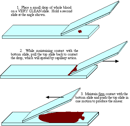
. On a blood smear slide prepared using Wrights stain you observe a large cell with a U-shaped nucleus and pale blue cytoplasm. It is imperative that proper tech nique be used in smear preparation. Move the spreader backward so that it makes contact with drop of blood.
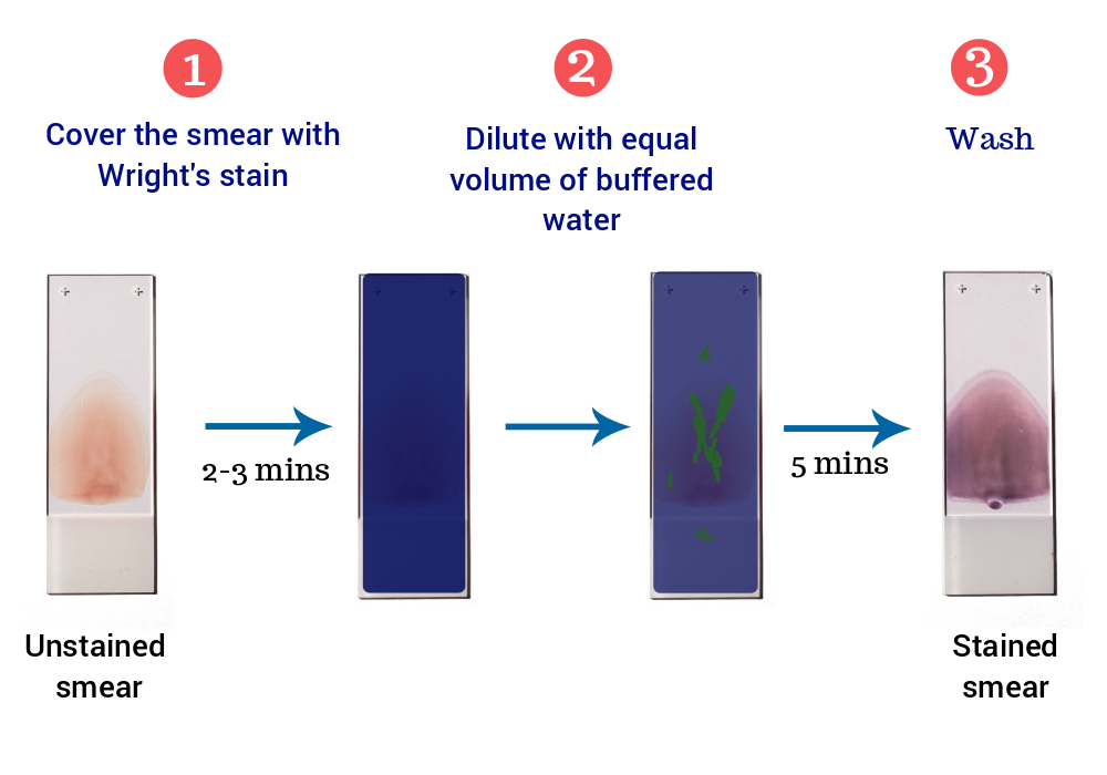
Place the air-dried smear on the slide staining rack smear side facing upwards. The stained smear will have no value and worse still. Once all set the next step is to add the buffer.
An unstained blood smear is clearly seen by eye on the microscope slide however the blood cells are barely visible under the microscope figure 1. Then move the spreader forward rapidly over the slide. Let stand for 2-3 minutes.
Megan Brashear CVT VTS ECC demonstrates the technique in staining a blood smear for microscopic evaluationbloodsmear veterinary veterinarymedicine-. Dry the slides upright in a rack. Dip the slide in methanol for a few seconds and ensure that it dries up completely.
Right blood as seen at 400x under light microscope. 1 thalassemia 2 leukocytosis 3 sickle-cell anemia 4 polycythemia. Wright - Geimsa and these stains give excellent result but tend to be fussy.
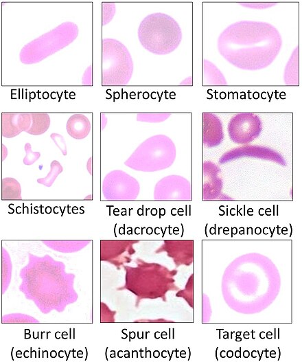
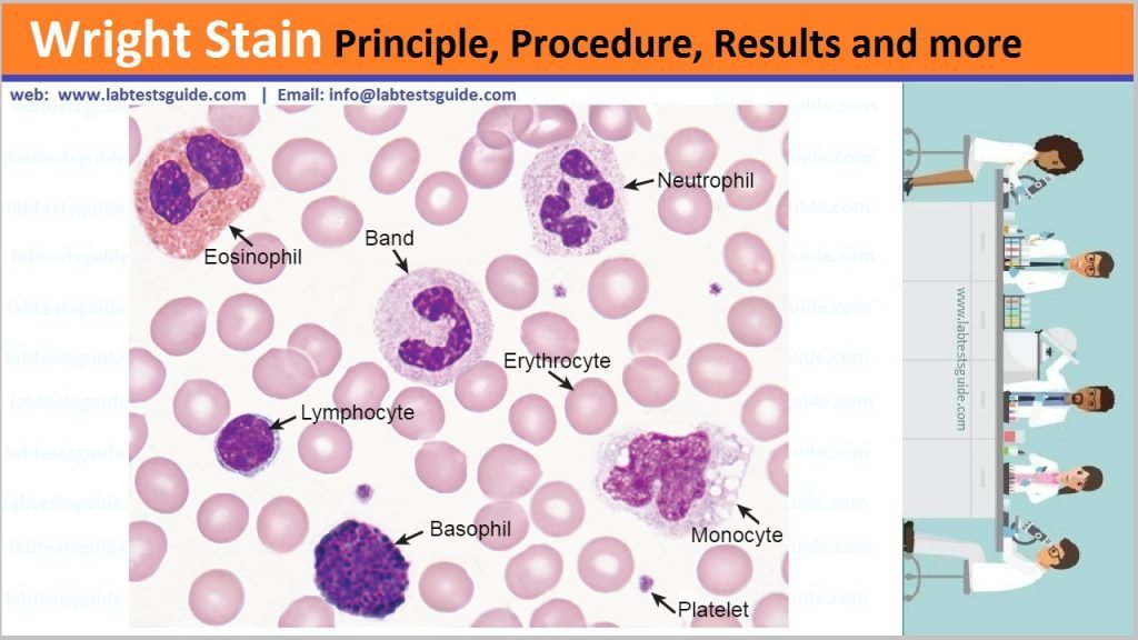
Cover the blood film with undiluted staining solution. Rinse slide or tape-strip in distilled water or Weises buffer pH 72. Slide staining Romanowsky-type stains give good nuclear and cytoplasmic detail.
The coverglass method and the slide method. Provide Content Correction We continue to work to improve your shopping experience and your feedback regarding this content is very important to us. Dip slide or tape-strip five times for one second each into Fixative.
Please use the form below to provide feedback related to the content on this product. Ideal for biology classrooms to explore structure-function relationships as per NGSS standards. On a blood smear slide prepared using Wrights stain you observe a large cell with a U-shaped nucleus and pale blue cytoplasm.
Place another slide spreader with smooth edge at an angle of 30-45⁰ near the drop of blood. The blood smear is stained with Wrights stain allowing students to easily distinguish between red and white blood cells. Human blood slide Wrights stain smear.
A thin peripheral blood film is thus prepared Dry it and. Drennan 1991 1 Prepare a solution of 20 ml Giemsa stain 240 ml deionized H2O. 1pcs Microscope Slides size.
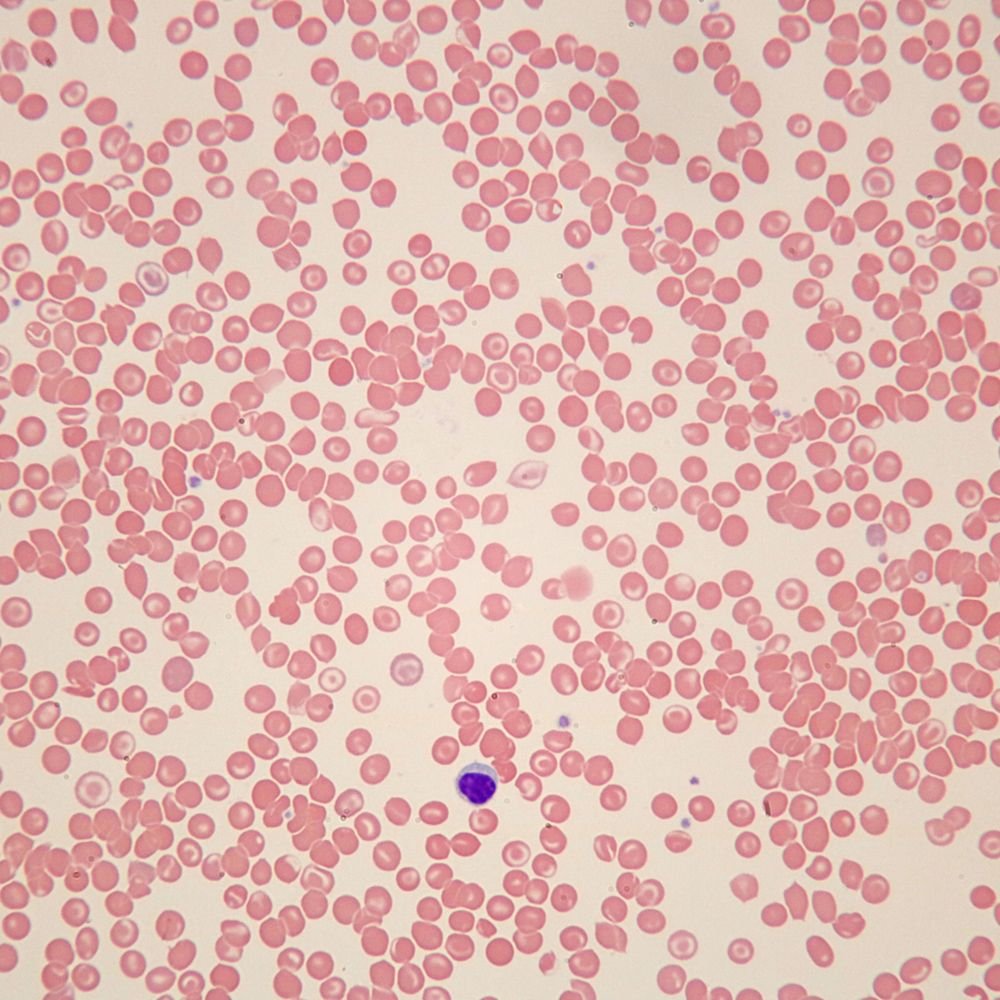
The undiluted stain fixes and partially stains the smear. Red blood cells stain red-orange nuclei stain blue-purple and cytoplasm stains blue to pink. 13 There are 2 procedures for making blood smears.
Wait for 8-10 minutes for staining to complete. Protected from light sample permanent storage. Professionally stained samples for best visualization.
Slotted slide case easy to storage. Dip slide or tape-strip five times for one second each into Stain 1. Prepare a film of blood or bone marrow on a microscopic slide and allow to air dry.
Making the Blood Smear Before any stained smear can be used for a diagnosis. Add 20 ml distilled water or Phosphate buffer pH 65 and let stand twice as long as 1-3 minuts. 1 x 3 254x762mm.
See delivery options in cart. Smear of human blood showing both erythrocytes red blood cells and different types of leukocytes white. Place slide on a level staining rack and place 10 ml of the Wright Stain Solution upon the smear 1 3 minutes.
One alternate is 10 minutes in 10 Giemsa. Mix the stain with water by blowing air with the help of a glass tube or with a dropper. The shorter stains yield faster.
1 monocyte 2 basophil 3 eosinophil 4 lymphocyte. Shows three main blood cell types - red blood cells white blood cells and. We also supply the following kinds of prepared microscope slides for biology science education and provide custom services.
Slide Method Procedure Place a drop of blood in the centre of a clean glass slide 1 to 2 cm from one end. The Wright stain also known as the Romanowsky stain is a mix of both. Talk to Customer Service.
Wrights stain procedure. 406-256-0990 or Live Chat In. This must be made fresh daily.
This cell is most likely a n __________. 2 Using a Beral pipette overlay a properly prepared blood smear slide with enough Wrights stain to completely cover the slide with a layer of stain approximately 18 thick. Begin the procedure with the fixative step.
Pre-cleaned glass slide with ground edges. This cell is most likely an _____. Place 3-4 good slides horizontally on the stand.
Give your students an up-close look at human blood cells using Carolinas most popular prepared blood microscope slide. Method Allow smears to dry. Ages 8 In Stock Ready to Ship Need it fast.
After one minute staining with black Quink the walls of the red blood cells erythrocytes. Leukocytes white blood cells are larger cells with a purple. The procedure involves taking a blood sample creating a smear using the wedge method most commonly then applying a stain.
Dip slide for a few seconds in methanol as a fixative step and allow slide to air dry completely. A basophil b lymphocyte c monocyte d eosinophil. As alternates to this 45-60 minutes in 25 Giemsa stain the smears could be stained for shorter times in more concentrated stains.
Remove thin smear slides and rinse by dipping 3-4 times in the Giemsa buffer. Thick smears should be left in buffer for 5 minutes. Dip slide or tape-strip five times for one second each into Stain 2.
May be seriously misleading. Left - unstained blood smear on slide. High quality optical glass.
Once the smear is air-dried completely place it on the slide staining rack and apply 1 ml of Wrights stain solution for about 3 minutes. Erythrocytes red blood cells are the plentiful pink cells in the smear.

Blood Smear Preparation And Staining Practical Lab Youtube
Blood Cytology
In Clinic Hematology The Blood Film Review Today S Veterinary Practice
Blood Smear Wikiwand

Eisco Prepared Microscope Slide Human Blood Smear Wright S Stain Microbiology Fisher Scientific

Blood Smear Wikiwand
Wright Stain Principle Procedure Result

Prepared Individual Microscope Slides Human Blood Smear Wright S Stain Education Fisher Scientific

Peripheral Blood 1000x Smear Wright S Stain Petroarc International
Wright S Stain Preparation Principle Procedure And Results

Peripheral Blood Smears From A Sprague Dawley Rat A Fresh Blood Download Scientific Diagram

Peripheral Blood Smears From A Sprague Dawley Rat A Fresh Blood Download Scientific Diagram
Pbs Prepararion Using Wrights Stain

Lecture Notes In Medical Technology Lecture 13 Morphological Examination Of Blood Films

Blood Smear Preparation And Staining

Blood Smear Wikiwand
Human Sickle Cell Anemia Slide Smear Wright S Stain Blood Smears Amazon Com Industrial Scientific
Wright Stain Principle Procedure Results And More Lab Tests Guide

Human Blood Film Slide Smear Wright S Stain Carolina Com


Comments
Post a Comment